Clinical Education
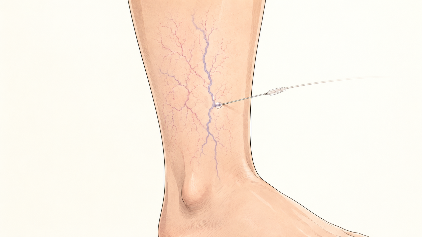
Sclerotherapy uses a very fine needle to deliver a small amount of medication into the target vein. The treated vessel gradually closes and fades. The vein does not disappear immediately; improvement usually develops over several weeks or months, and more than one treatment session may be needed.
- Guideline recommendation: Liquid or foam sclerotherapy may be considered for symptomatic spider veins or reticular veins.
- Research finding: Both sclerotherapy and laser treatment can improve small leg veins, but a stronger medication is not automatically better.
- Dr LiangVascular’s clinical interpretation: Assess the vessel pattern and feeding veins first, then use an appropriate concentration, small amounts and a staged approach.
How do spider veins, reticular veins and bulging varicose veins differ?
- Spider veins usually appear as fine red or purple lines near the skin surface. Reticular veins are generally slightly larger and blue-green, while bulging varicose veins are larger, twisted vessels that may project above the skin.
- Appearance helps describe the type of visible vessel, but it cannot show where reflux begins or determine by itself whether treatment is needed.
- Assessment still considers symptoms, swelling or skin change, vessel distribution and clinical examination. Standing leg-vein ultrasound is considered only when clinically appropriate.
Do spider veins alone need medical assessment?
- A small number of spider veins without aching, swelling, skin change or larger varicose veins does not automatically require treatment or a routine full duplex scan.
- Assessment may be helpful when appearance is a concern, the vessel pattern is increasing, or there are associated symptoms, one-sided swelling, skin changes, larger varicose veins or broader clinical concerns.
- Observation, further investigation and treatment should be selected according to the individual clinical context and the patient's goals.
Treatment is not simply injecting every visible vein
- Spider veins may be connected to slightly larger feeding veins beneath the skin. Treating only the visible surface vessels without identifying these connections may produce an incomplete result or allow the veins to reappear sooner.
- Before treatment, I assess the colour, size, depth and pattern of the vessels. These findings help determine the injection order, medication concentration and area treated during each session.
- The goal is not to perform as many injections as possible. It is to use an appropriate technique and work gradually from the larger feeding vessels towards the finer spider veins.
What reactions can occur after treatment?
- Common reactions include bruising, local tenderness, temporary pigmentation and the appearance of very fine new vessels around the treated area.
- Less common complications include skin injury, thrombosis and allergic reactions.
- A 2023 systematic review found that both sclerotherapy and laser treatments can improve small leg veins. However, a stronger sclerosant is not automatically better, because increasing potency may also increase unwanted effects such as pigmentation.
How many sessions are needed?
- Spider veins often require patience and staged treatment. The number of sessions depends on the number, size and depth of the vessels, as well as the skin response.
- After each treatment, the vessels need time to fade before the next stage is assessed. Even after successful treatment, new small vessels may still develop in the future.
Four things to assess before sclerotherapy
| Assessment | Why it matters | Common approach |
|---|---|---|
| Vessel size and depth | Affects medication concentration and injection order | Work gradually from larger feeding veins |
| Aching or swelling | May indicate more than a cosmetic concern | Arrange an ultrasound when appropriate |
| Skin response | Bruising and pigmentation need time to improve | Observe between stages |
| Treatment expectations | Spider veins do not all disappear after one session | Discuss likely sessions and recovery time |
Frequently asked questions
Are spider veins the same as bulging varicose veins?
No. Spider veins are fine red or purple surface vessels, while bulging varicose veins are generally larger and twisted. Appearance alone cannot identify the source of reflux.
Do a few spider veins always require standing duplex ultrasound?
No. A routine full scan may not be necessary when there are no symptoms, swelling, skin changes or larger varicose veins. The decision remains clinical.
Can spider veins be treated directly with sclerotherapy?
The vessel type, distribution, associated symptoms and treatment goal should be assessed first. Sclerotherapy may be considered for selected local vessels, but not every visible vein should be injected automatically.
Note: Sudden one-sided swelling, warmth, significant pain or shortness of breath requires prompt medical attention. These symptoms should not be treated as a cosmetic concern alone.
References
- Gloviczki P, Lawrence PF, Wasan SM, et al. The 2023 Society for Vascular Surgery, American Venous Forum, and American Vein and Lymphatic Society Clinical Practice Guidelines for the Management of Varicose Veins of the Lower Extremities. Part II. Journal of Vascular Surgery: Venous and Lymphatic Disorders. 2024;12(1):101670. View source
- De Maeseneer MG, Kakkos SK, Aherne T, et al. European Society for Vascular Surgery 2022 Clinical Practice Guidelines on the Management of Chronic Venous Disease of the Lower Limbs. European Journal of Vascular and Endovascular Surgery. 2022;63(2):184–267. View source
- Bontinis V, Bontinis A, Koutsoumpelis A, et al. Interventions for the Treatment of Lower Limb Telangiectasias and Reticular Veins: A Systematic Review and Network Meta-Analysis. European Journal of Vascular and Endovascular Surgery. 2023;66(4):560–576. View source
- Tan M, Shaydakov E, Parsi K, Davies AH; International Union of Phlebology. Microsclerotherapy. Phlebology. 2024;39(4):280–283. View source
This article is for health education only and does not replace individual medical assessment. Seek urgent care for acute symptoms.
Dr LiangVascular
Begin with a careful assessment.
Symptoms, examination and ultrasound findings are considered before treatment options are discussed.
Book an Appointment