Clinical Education
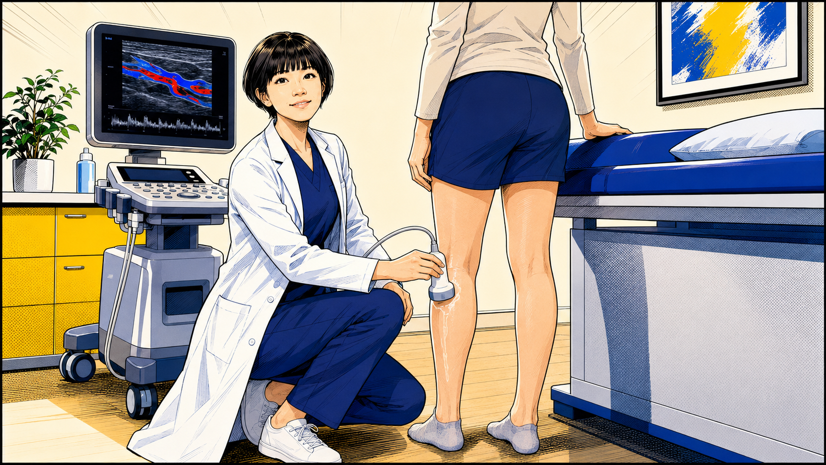
The examination is non-invasive and uses no radiation or injections. According to the clinical situation, the veins are assessed while standing or in another dependent position that allows adequate venous filling, with manoeuvres used to observe blood flow.
- Maps the location, direction and extent of reflux when clinically appropriate
- Assesses the superficial and deep venous systems
- Supports diagnosis and treatment planning but does not decide treatment by itself
When might standing venous duplex ultrasound be needed?
- Symptomatic varicose veins, including aching, heaviness, swelling or itching, and varicose veins that have appeared again after previous treatment.
- Swelling, skin discoloration or induration, or a wound that is not healing normally when venous disease is clinically suspected.
- Treatment planning when venous anatomy, the source of reflux and connections between vessels need clarification.
- Persistent symptoms, appearance concerns or new venous changes after previous treatment, when residual, recurrent or newly developed reflux needs to be distinguished.
When might a routine full scan not be necessary?
- A small number of asymptomatic superficial vessels that are purely cosmetic does not automatically require a routine full duplex examination.
- The value of scanning changes when there are associated symptoms, swelling, skin changes, larger varicose veins, recurrent vessels or broader clinical concerns.
- The decision remains based on the history, symptoms, examination and the problem the patient wants assessed.
What does the examination show?
- The anatomy, patency and blood-flow direction of the superficial and deep venous systems.
- The location, direction and extent of reflux, and the pattern of connections between related veins.
- Residual, recurrent or newly developed reflux after previous treatment.
- A venous map that can support treatment planning when intervention is being considered.
What can ultrasound not decide by itself?
- A vein diameter or an isolated reflux finding should not by itself determine whether treatment is necessary.
- Reflux on ultrasound does not prove that every episode of leg pain, swelling or other discomfort is caused by that vein.
- Symptoms, examination, anatomy, reflux pattern, skin changes, impact on daily life and individual circumstances must be interpreted together.
How should I prepare?
- Fasting is usually unnecessary, and the examination uses no radiation or contrast injection.
- Wear clothing that allows the thigh and lower leg to be exposed, and bring previous ultrasound, procedure or treatment records.
- Note where and when symptoms occur, whether they relate to standing or activity, and the questions you most want answered.
Appearance alone compared with duplex assessment
| Comparison | Looking at the leg | Standing duplex ultrasound |
|---|---|---|
| Information | Surface appearance | Superficial and deep veins, blood-flow direction and reflux pattern |
| Source of reflux | Cannot be identified reliably | Can be mapped when clinically appropriate |
| Treatment planning | May focus only on visible vessels | Helps integrate anatomy, source and treatment extent |
| Examination | Visual and physical examination | Non-invasive, no radiation and usually no injection |
Frequently asked questions
Does every first vascular appointment include standing duplex ultrasound?
No. The history, symptoms and clinical examination come first. Duplex is arranged when a venous cause is suspected or when reflux mapping, recurrent-disease assessment or treatment planning is clinically useful.
Why is venous ultrasound often performed while standing or in a dependent position?
Allowing the leg veins to fill under gravity helps the examiner observe blood flow and reflux. The exact position is adjusted for safety, the patient's condition and the purpose of the examination.
Does a larger vein or reflux on ultrasound mean treatment is necessary?
No. A vein diameter or isolated reflux finding should not determine treatment by itself. Symptoms, examination, anatomy, reflux pattern, skin changes, impact on daily life and individual circumstances must be considered together.
Note: Not every small visible vein requires a full duplex scan, and treatment should not be decided from a single ultrasound measurement. Symptoms, clinical findings and individual circumstances remain essential.
References
- Gloviczki P, Lawrence PF, Wasan SM, et al. The 2022 Society for Vascular Surgery, American Venous Forum, and American Vein and Lymphatic Society Clinical Practice Guidelines for the Management of Varicose Veins of the Lower Extremities. Part I: Duplex Scanning and Treatment of Superficial Truncal Reflux. Journal of Vascular Surgery: Venous and Lymphatic Disorders. 2023;11(2):231–261.e6. View source
- De Maeseneer MG, Kakkos SK, Aherne T, et al. European Society for Vascular Surgery 2022 Clinical Practice Guidelines on the Management of Chronic Venous Disease of the Lower Limbs. European Journal of Vascular and Endovascular Surgery. 2022;63(2):184–267. View source
- National Institute for Health and Care Excellence. Varicose veins: diagnosis and management (CG168). Duplex ultrasound assessment recommendations. View source
This article is for health education only and does not replace individual medical assessment. Seek urgent care for acute symptoms.
Dr LiangVascular
Begin with a careful assessment.
Symptoms, examination and ultrasound findings are considered before treatment options are discussed.
Book an Appointment