Clinical Education
Varicose vein treatment is therefore not simply a matter of treating each visible vein. I first use standing leg-vein ultrasound to locate the source of reflux, then plan how the truncal vein and visible branches should be managed.
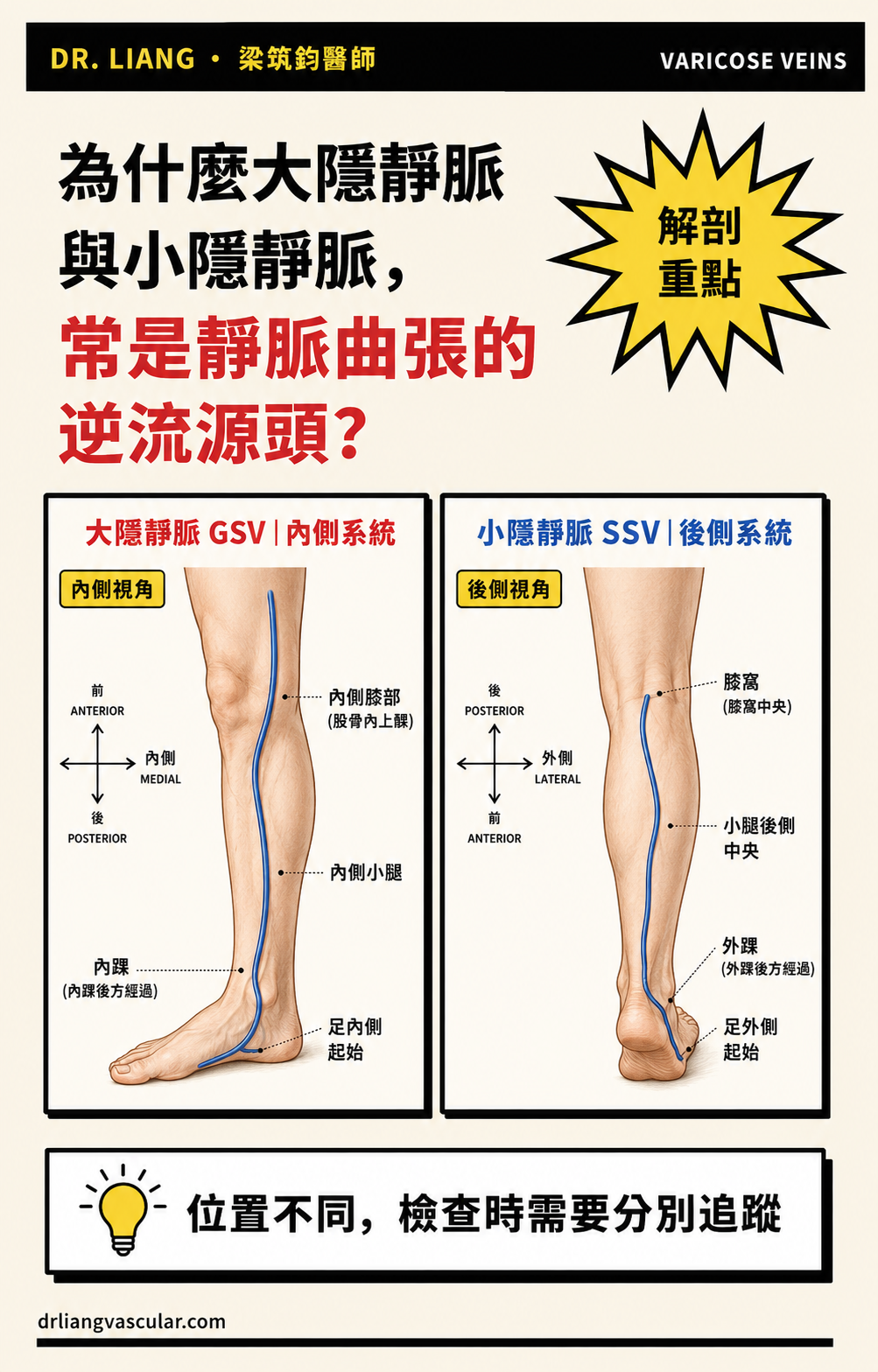
- The great saphenous vein (GSV) and small saphenous vein (SSV) are major superficial venous trunks
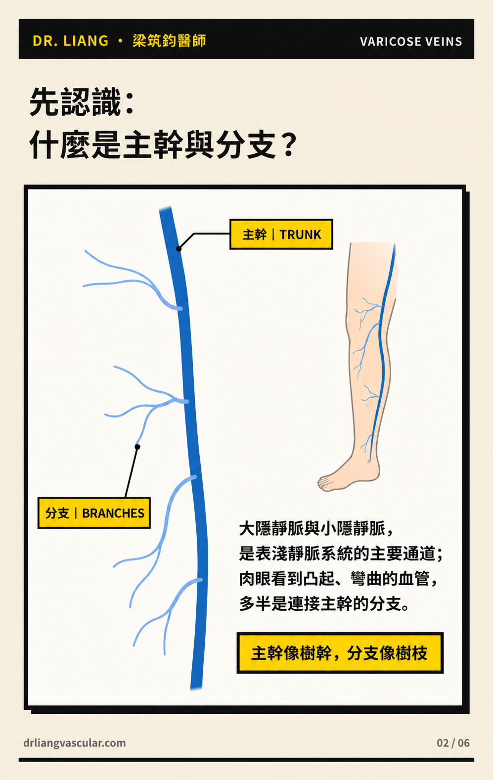
- Bulging, tortuous veins seen on the skin are often tributaries
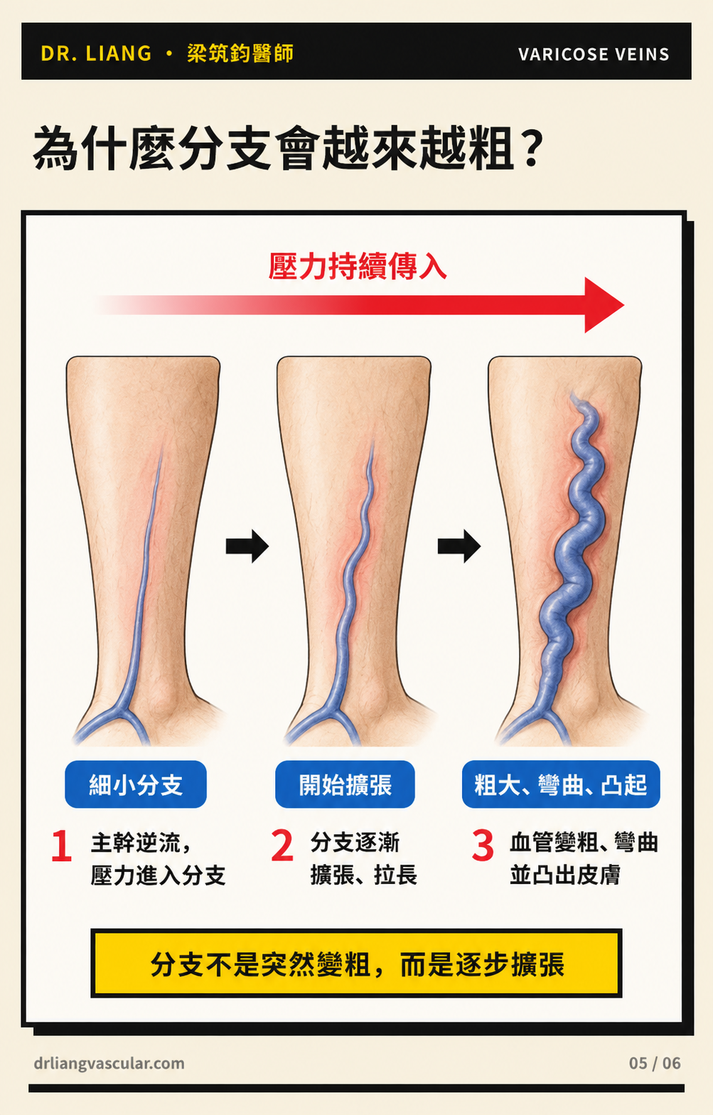
- When valves fail, pressure can travel through the trunk and progressively enlarge connected branches
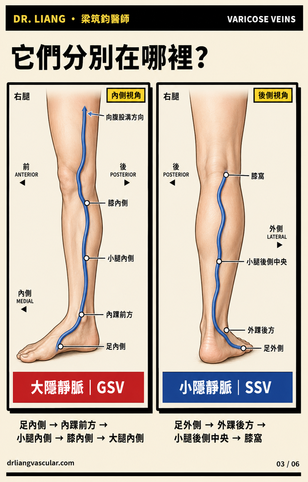
Start by separating trunks from branches
- The GSV begins on the medial side of the foot, passes in front of the medial ankle, and travels along the medial calf, knee and thigh.
- The SSV begins on the lateral side of the foot, passes behind the lateral ankle, and usually travels up the posterior calf.
- The rope-like veins visible under the skin are often tributaries connected to these trunks rather than the trunks themselves.
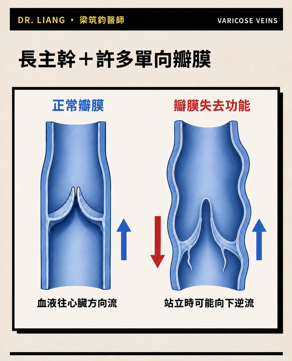
Venous valves work like one-way doors
- Healthy valves work with the calf-muscle pump to move blood toward the heart.
- When a valve does not close properly, blood can flow downward while standing and increase pressure in the trunk.
- Surface appearance cannot identify which valve segment has failed; ultrasound is needed to map reflux.
Why do tributaries become larger?
- Persistent truncal reflux can transmit pressure to connected tributaries.
- Over time, these branches may dilate, lengthen and become tortuous before they appear as visible bulging veins.
- This is usually a progressive process rather than a change that happens overnight.
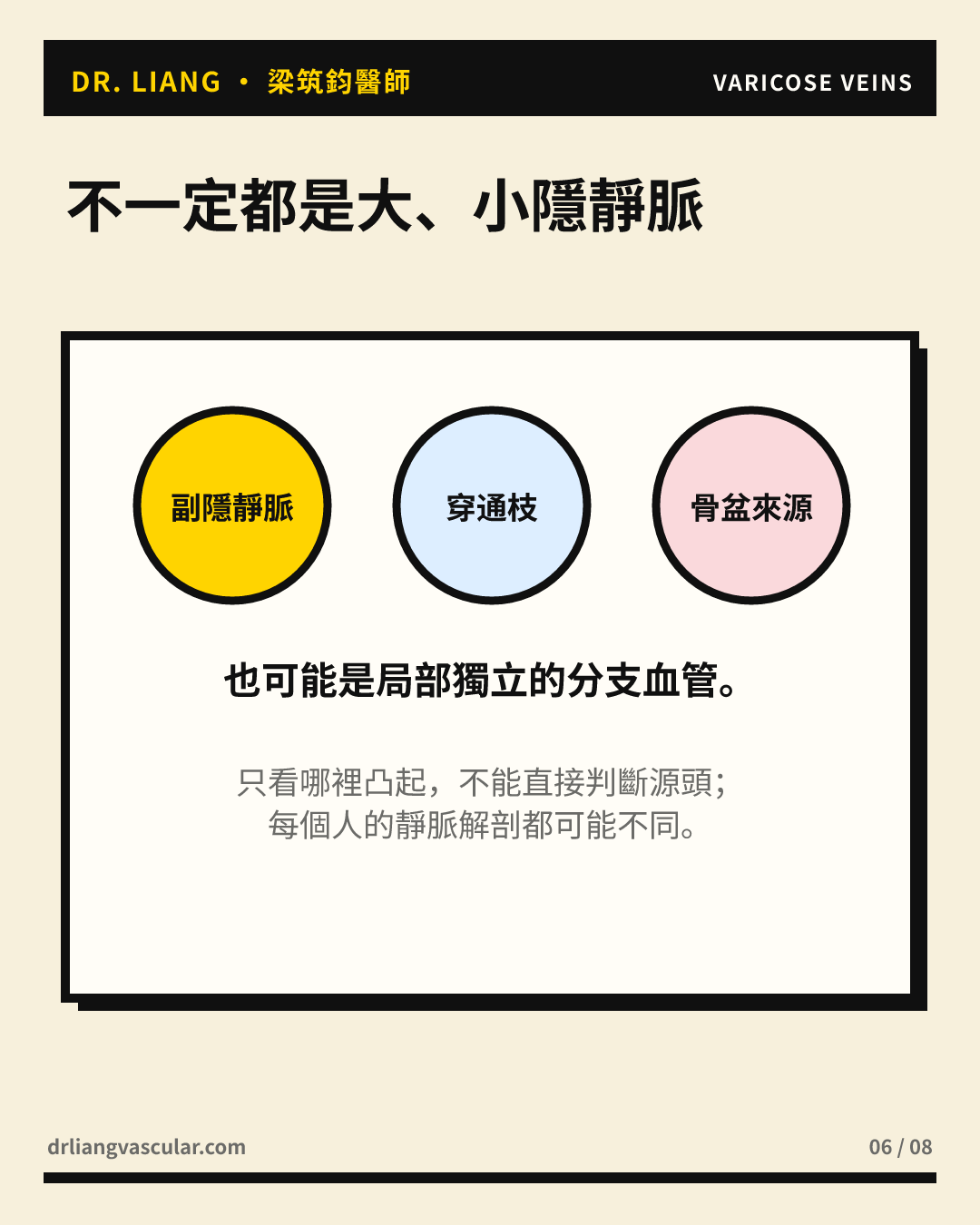
Not every visible vein has the same source
- Some tributaries are related to GSV or SSV reflux, while others may arise from accessory veins, perforators or a more local source.
- Two people with similar-looking veins may therefore need different treatment plans.
- My approach is to identify the source first, then decide whether visible branches should be treated at the same time or in stages.
Truncal veins and tributaries
| Comparison | GSV / SSV trunks | Superficial tributaries |
|---|---|---|
| Course | More predictable anatomical pathway | More variable pathways connected to trunks |
| Visibility | Not always visibly prominent | More often forms visible tortuous veins |
| Role | May be the source of reflux | May enlarge from transmitted pressure or have a separate source |
| Assessment | Standing leg-vein ultrasound | Clinical examination plus ultrasound mapping |
Frequently asked questions
Does a bulging vein mean that the GSV is always abnormal?
No. A visible vein is often a tributary and may connect to the GSV, SSV, an accessory vein, a perforator or another local source. Ultrasound is needed to confirm the anatomy.
Will visible branches disappear after truncal treatment?
Some branches become smaller, but not all disappear. Microphlebectomy or sclerotherapy may still be appropriate, either at the same session or later, depending on vein size, symptoms and personal goals.
How can the source of reflux be identified?
Standing leg-vein ultrasound maps blood-flow direction, the extent of reflux, and the relationship between superficial trunks, tributaries and deep veins.
Note: Seek prompt medical care for sudden one-sided leg swelling or pain, redness and heat, or active bleeding from a varicose vein.
References
- De Maeseneer MG, Kakkos SK, Aherne T, et al. European Society for Vascular Surgery (ESVS) 2022 Clinical Practice Guidelines on the Management of Chronic Venous Disease of the Lower Limbs. Eur J Vasc Endovasc Surg. 2022;63(2):184–267. doi:10.1016/j.ejvs.2021.12.024. View source
- Gloviczki P, Lawrence PF, Wasan SM, et al. The 2022 Society for Vascular Surgery, American Venous Forum, and American Vein and Lymphatic Society clinical practice guidelines for the management of varicose veins of the lower extremities. Part I. J Vasc Surg Venous Lymphat Disord. 2023;11(2):231–261.e6. doi:10.1016/j.jvsv.2022.09.004. View source
- Gloviczki P, Lawrence PF, Wasan SM, et al. The 2023 Society for Vascular Surgery, American Venous Forum, and American Vein and Lymphatic Society clinical practice guidelines for the management of varicose veins of the lower extremities. Part II. J Vasc Surg Venous Lymphat Disord. 2024;12(1):101670. doi:10.1016/j.jvsv.2023.08.011. View source
- Caggiati A, Bergan JJ, Gloviczki P, et al. Nomenclature of the veins of the lower limb: extensions, refinements, and clinical application. J Vasc Surg. 2005;41(4):719–724. doi:10.1016/j.jvs.2005.01.018. View source
This article provides general health education and does not replace an individual medical assessment. The source of reflux, venous anatomy and treatment plan should be evaluated with standing ultrasound and your clinical symptoms.
Dr LiangVascular
Begin with a careful assessment.
Symptoms, examination and ultrasound findings are considered before treatment options are discussed.
Book an Appointment