中文版
因此,治療靜脈曲張不是看到一條、處理一條。我會先用站立式下肢靜脈超音波,找出逆流從哪裡開始,再決定主幹與分支要如何搭配處理。
- 大隱靜脈(GSV)與小隱靜脈(SSV)是表淺靜脈系統的重要主幹
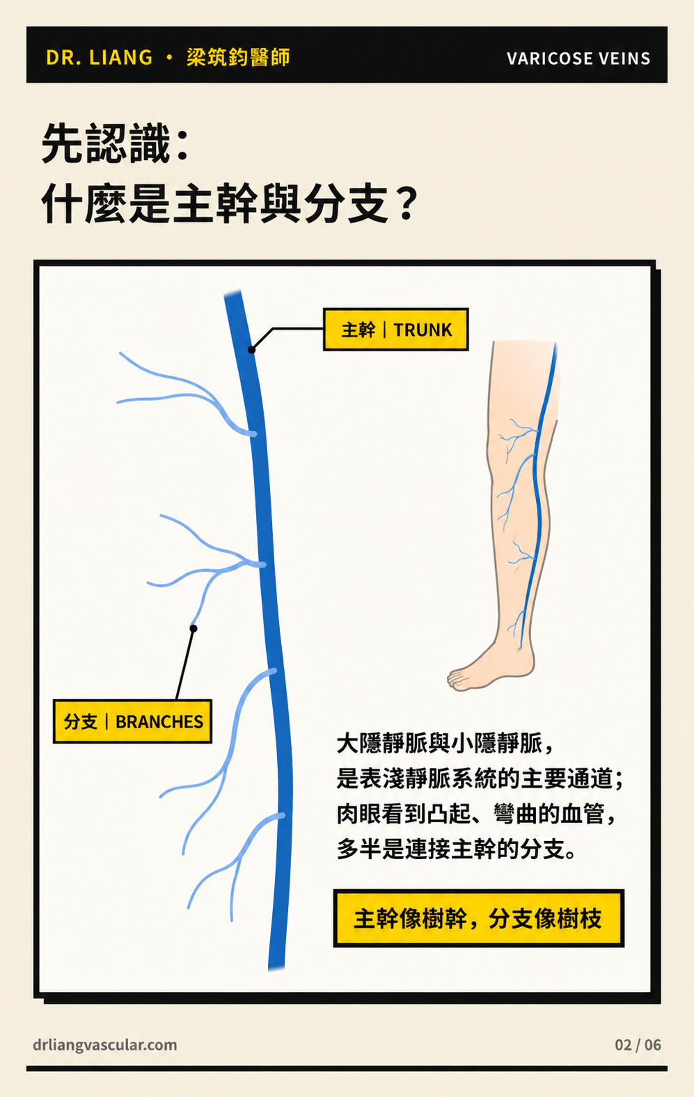
- 肉眼看到凸起、彎曲的血管,常是連接主幹的分支
- 瓣膜關不緊時,壓力可能沿主幹傳到分支,讓分支逐漸擴張
先分清楚主幹與分支
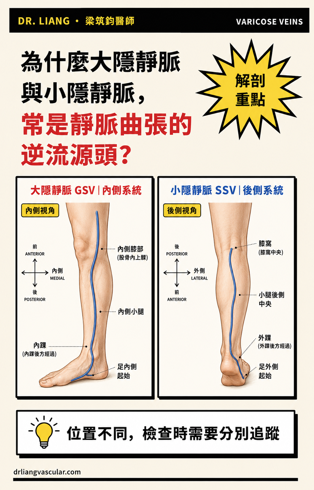
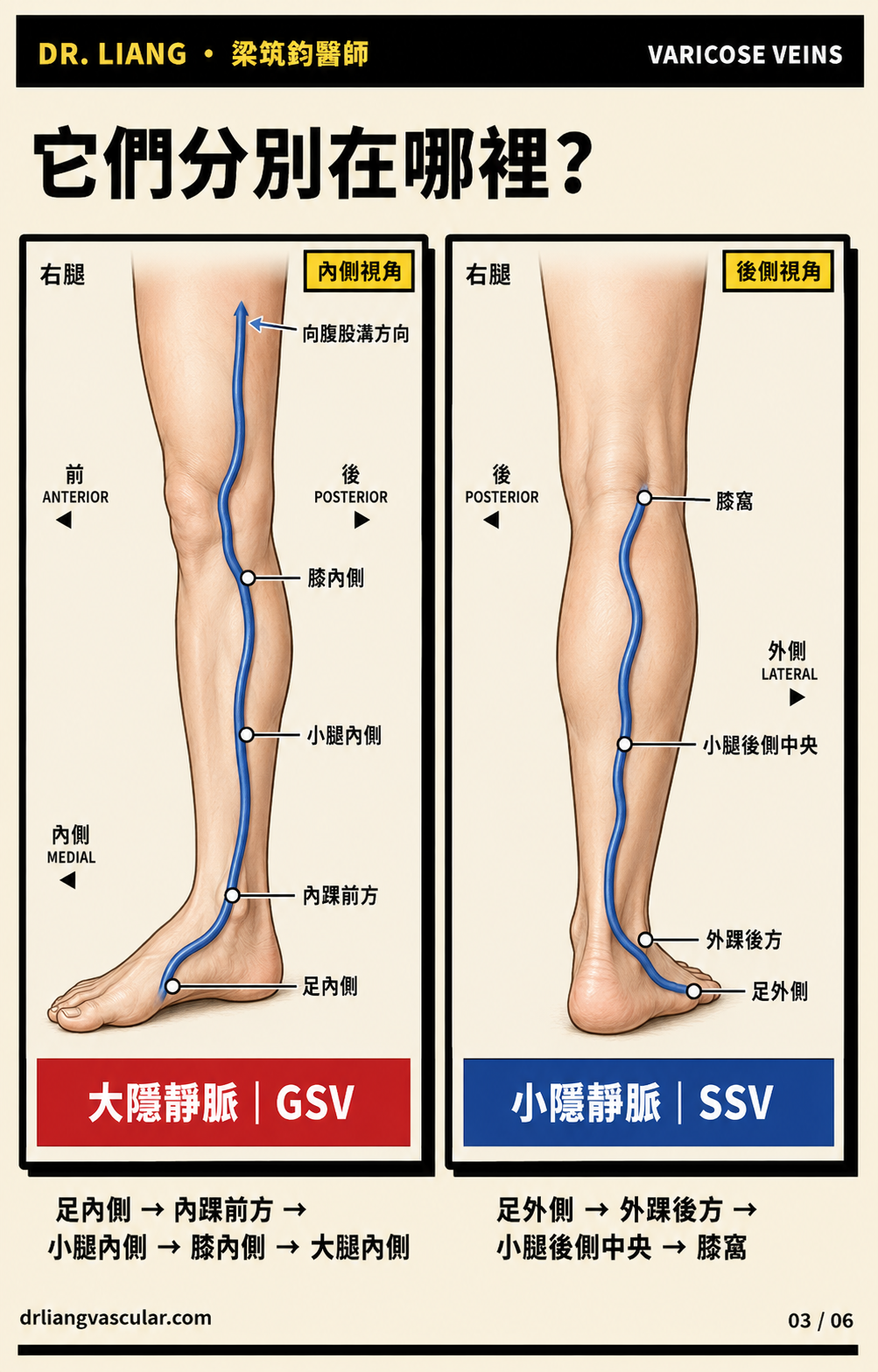
- 大隱靜脈從足部內側開始,經過內踝前方,沿小腿、膝部與大腿內側向上走。
- 小隱靜脈從足部外側開始,經過外踝後方,主要沿小腿後側向上走。
- 腿上肉眼看得到、像繩子一樣凸起的血管,很多是接在主幹上的分支,不一定就是主幹本身。
瓣膜像血管裡的單向門
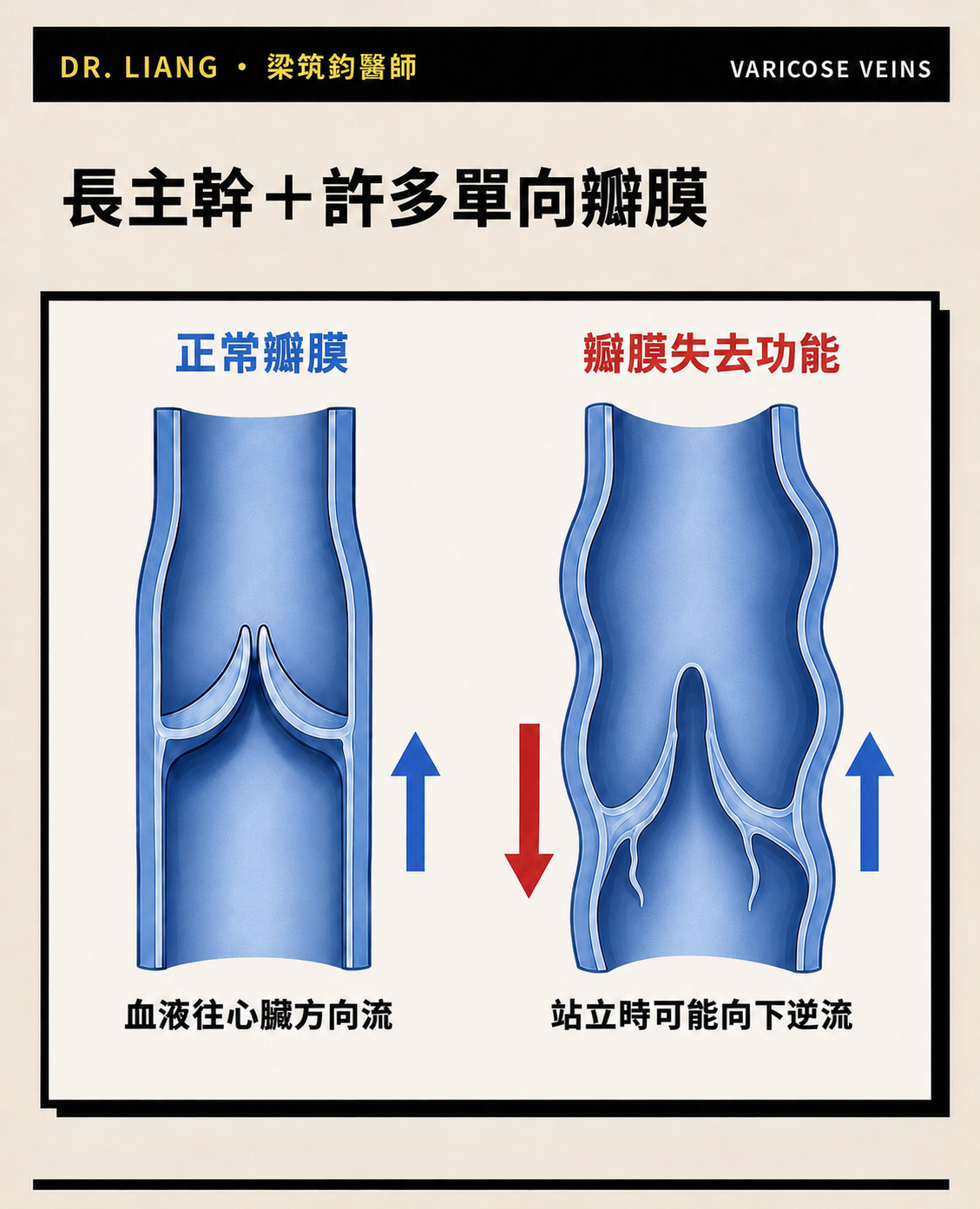
- 正常情況下,瓣膜會配合小腿肌肉收縮,幫助血液往心臟方向走。
- 當瓣膜關不緊,站立時血液可能往下逆流,主幹內的壓力就會增加。
- 外觀只能看到表面,無法判斷哪一段瓣膜失去功能,所以需要超音波確認。
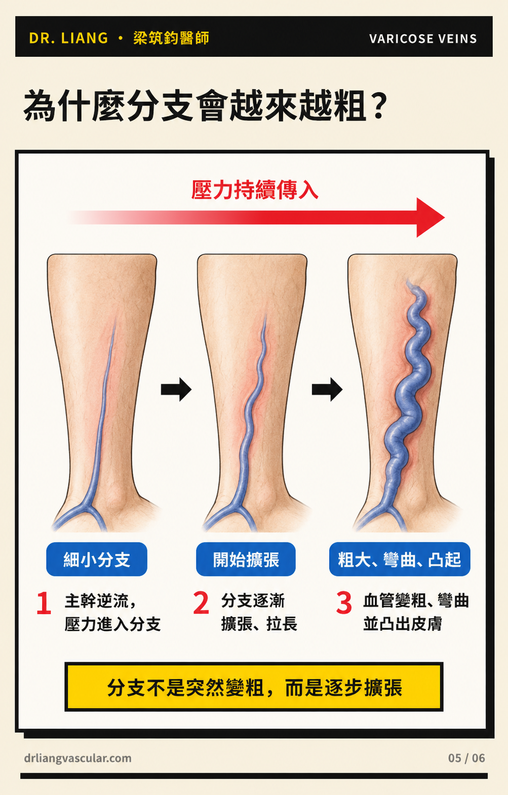
為什麼分支會越來越粗?
- 如果主幹持續逆流,壓力可能傳到相連的分支。
- 分支長期承受壓力後,會慢慢擴張、拉長、彎曲,最後變成肉眼可見的凸起血管。
- 這通常是逐漸發生的過程,不是某一天突然長出來。
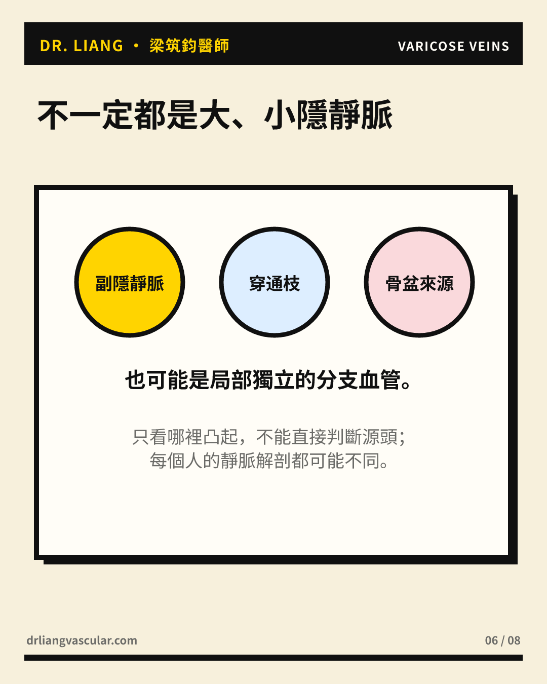
不是每一條凸起血管都來自同一個主幹
- 有些分支與大隱或小隱靜脈逆流有關,有些則可能來自副隱靜脈、穿通枝或局部獨立的逆流。
- 因此,同樣是腿上凸起的血管,每個人的來源與治療方式都可能不同。
- 我的處理原則是先找逆流源頭,再評估可見分支是否需要同時或分階段處理。
主幹與分支,差別在哪裡?
| 比較 | 大隱/小隱靜脈主幹 | 表淺分支 |
|---|---|---|
| 位置與走向 | 有較固定的解剖路徑 | 走向較多變,與主幹相連 |
| 外觀看得到嗎? | 不一定明顯凸起 | 較常形成肉眼可見的彎曲血管 |
| 在靜脈曲張中的角色 | 可能是逆流的源頭 | 可能因壓力而擴張,也可能有獨立來源 |
| 如何確認 | 站立式下肢靜脈超音波 | 外觀檢查加上超音波追查來源 |
常見問題
腿上有凸起血管,就代表大隱靜脈一定異常嗎?
不一定。凸起血管常是分支,可能連接大隱靜脈、小隱靜脈、副隱靜脈、穿通枝或其他局部來源,需要超音波確認。
主幹治療後,凸起的分支會自己消失嗎?
部分分支可能縮小,但不一定全部消失。有些仍需要微創切除或硬化劑注射,是否同時處理或分階段處理,要依血管大小、症狀與個人需求決定。
要怎麼知道逆流真正從哪裡開始?
最實用的方法是站立式下肢靜脈超音波。檢查會追蹤血液方向、逆流範圍,以及主幹、分支與深部靜脈的狀況。
提醒:如果單側腿突然腫痛、發紅發熱,或曲張血管正在出血,請儘快就醫,不要只靠外觀自行判斷。
English
Varicose vein treatment is therefore not simply a matter of treating each visible vein. I first use standing leg-vein ultrasound to locate the source of reflux, then plan how the truncal vein and visible branches should be managed.
- The great saphenous vein (GSV) and small saphenous vein (SSV) are major superficial venous trunks
- Bulging, tortuous veins seen on the skin are often tributaries
- When valves fail, pressure can travel through the trunk and progressively enlarge connected branches
Start by separating trunks from branches
- The GSV begins on the medial side of the foot, passes in front of the medial ankle, and travels along the medial calf, knee and thigh.
- The SSV begins on the lateral side of the foot, passes behind the lateral ankle, and usually travels up the posterior calf.
- The rope-like veins visible under the skin are often tributaries connected to these trunks rather than the trunks themselves.
Venous valves work like one-way doors
- Healthy valves work with the calf-muscle pump to move blood toward the heart.
- When a valve does not close properly, blood can flow downward while standing and increase pressure in the trunk.
- Surface appearance cannot identify which valve segment has failed; ultrasound is needed to map reflux.
Why do tributaries become larger?
- Persistent truncal reflux can transmit pressure to connected tributaries.
- Over time, these branches may dilate, lengthen and become tortuous before they appear as visible bulging veins.
- This is usually a progressive process rather than a change that happens overnight.
Not every visible vein has the same source
- Some tributaries are related to GSV or SSV reflux, while others may arise from accessory veins, perforators or a more local source.
- Two people with similar-looking veins may therefore need different treatment plans.
- My approach is to identify the source first, then decide whether visible branches should be treated at the same time or in stages.
Truncal veins and tributaries
| Comparison | GSV / SSV trunks | Superficial tributaries |
|---|---|---|
| Course | More predictable anatomical pathway | More variable pathways connected to trunks |
| Visibility | Not always visibly prominent | More often forms visible tortuous veins |
| Role | May be the source of reflux | May enlarge from transmitted pressure or have a separate source |
| Assessment | Standing leg-vein ultrasound | Clinical examination plus ultrasound mapping |
Frequently asked questions
Does a bulging vein mean that the GSV is always abnormal?
No. A visible vein is often a tributary and may connect to the GSV, SSV, an accessory vein, a perforator or another local source. Ultrasound is needed to confirm the anatomy.
Will visible branches disappear after truncal treatment?
Some branches become smaller, but not all disappear. Microphlebectomy or sclerotherapy may still be appropriate, either at the same session or later, depending on vein size, symptoms and personal goals.
How can the source of reflux be identified?
Standing leg-vein ultrasound maps blood-flow direction, the extent of reflux, and the relationship between superficial trunks, tributaries and deep veins.
Note: Seek prompt medical care for sudden one-sided leg swelling or pain, redness and heat, or active bleeding from a varicose vein.
References
- De Maeseneer MG, Kakkos SK, Aherne T, et al. European Society for Vascular Surgery (ESVS) 2022 Clinical Practice Guidelines on the Management of Chronic Venous Disease of the Lower Limbs. Eur J Vasc Endovasc Surg. 2022;63(2):184–267. doi:10.1016/j.ejvs.2021.12.024. 查看來源
- Gloviczki P, Lawrence PF, Wasan SM, et al. The 2022 Society for Vascular Surgery, American Venous Forum, and American Vein and Lymphatic Society clinical practice guidelines for the management of varicose veins of the lower extremities. Part I. J Vasc Surg Venous Lymphat Disord. 2023;11(2):231–261.e6. doi:10.1016/j.jvsv.2022.09.004. 查看來源
- Gloviczki P, Lawrence PF, Wasan SM, et al. The 2023 Society for Vascular Surgery, American Venous Forum, and American Vein and Lymphatic Society clinical practice guidelines for the management of varicose veins of the lower extremities. Part II. J Vasc Surg Venous Lymphat Disord. 2024;12(1):101670. doi:10.1016/j.jvsv.2023.08.011. 查看來源
- Caggiati A, Bergan JJ, Gloviczki P, et al. Nomenclature of the veins of the lower limb: extensions, refinements, and clinical application. J Vasc Surg. 2005;41(4):719–724. doi:10.1016/j.jvs.2005.01.018. 查看來源
本文提供一般衛教資訊,不能取代個別診斷或治療。靜脈曲張的逆流來源、血管走向與治療方式需依站立式超音波及個人症狀評估。
Dr LiangVascular
從完整評估開始
先整合症狀、理學檢查與超音波結果,再討論適合的治療選擇。
預約門診