Clinical Education
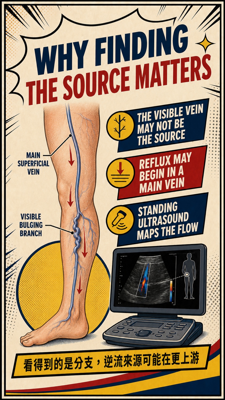
The veins you can see are often superficial tributaries, but they are not necessarily the source of reflux. Standing leg-vein ultrasound traces blood-flow direction and helps determine whether reflux arises from a main trunk, accessory vein, perforator or a more local branch.
- Similar-looking bulging veins can have very different sources of reflux
- Standing ultrasound maps blood-flow direction, source and extent
- Treatment should be planned according to symptoms, anatomy and ultrasound findings
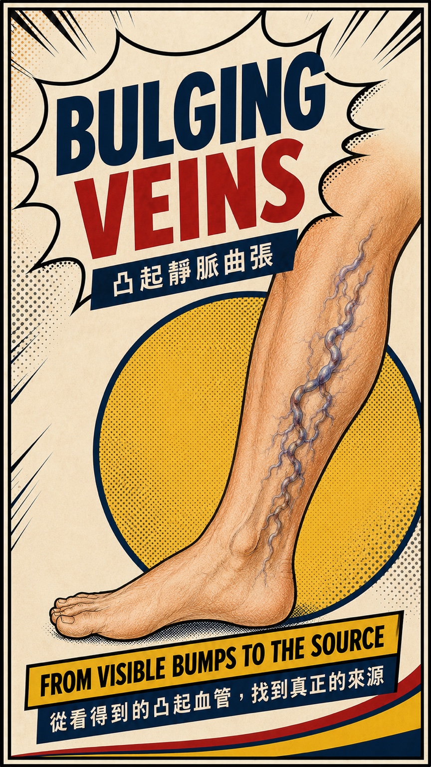
Bulging veins are more than an appearance issue
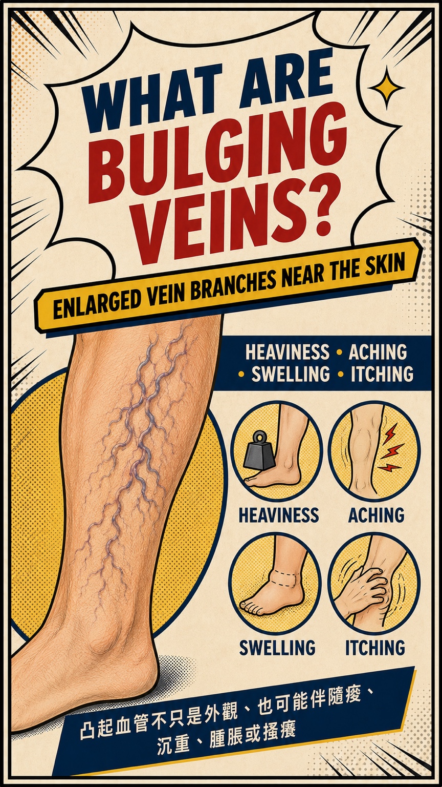
- Bulging varicose veins are enlarged, tortuous superficial branches located close to the skin.
- Some people have no significant discomfort, while others experience heaviness, aching, swelling, itching or symptoms that worsen after standing.
- Progressive ankle discoloration, firmness, recurrent inflammation or a slow-healing wound deserves earlier assessment.
Why does finding the source matter?
- The visible vein may be a downstream branch while reflux begins in a main trunk, accessory vein or perforator further upstream.
- In other people, the main trunks remain competent and the problem is mainly local. Appearance alone cannot determine the right treatment.
- Standing ultrasound traces the direction, starting point and extent of reflux to create an individual vein map.
What do I check?
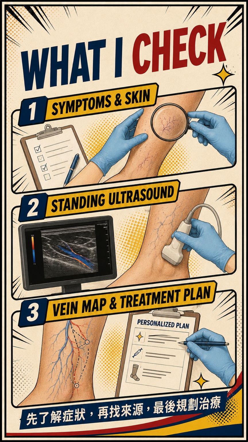
- We first discuss the symptoms that matter most, their timing, the effect of prolonged standing, previous treatment and skin changes.
- When appropriate, standing leg-vein ultrasound assesses the superficial and deep veins and traces the source of reflux.
- Symptoms, appearance, venous anatomy and ultrasound findings are then combined into a treatment sequence with realistic expectations.
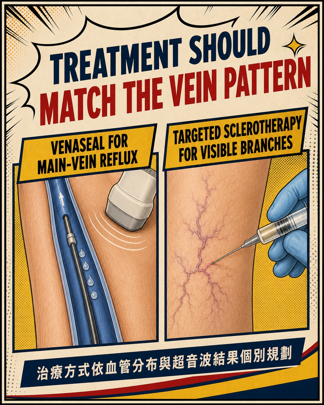
Treatment should match the vein pattern
- When suitable main-vein reflux is present, VenaSeal medical adhesive or another treatment may be considered according to anatomy, allergy history and personal needs.
- Visible bulging tributaries may be treated with ultrasound-guided sclerotherapy, microphlebectomy or another appropriate method.
- Trunks and branches may be treated together or in stages. Not every patient needs VenaSeal, and not every bulging vein is suitable for direct injection.
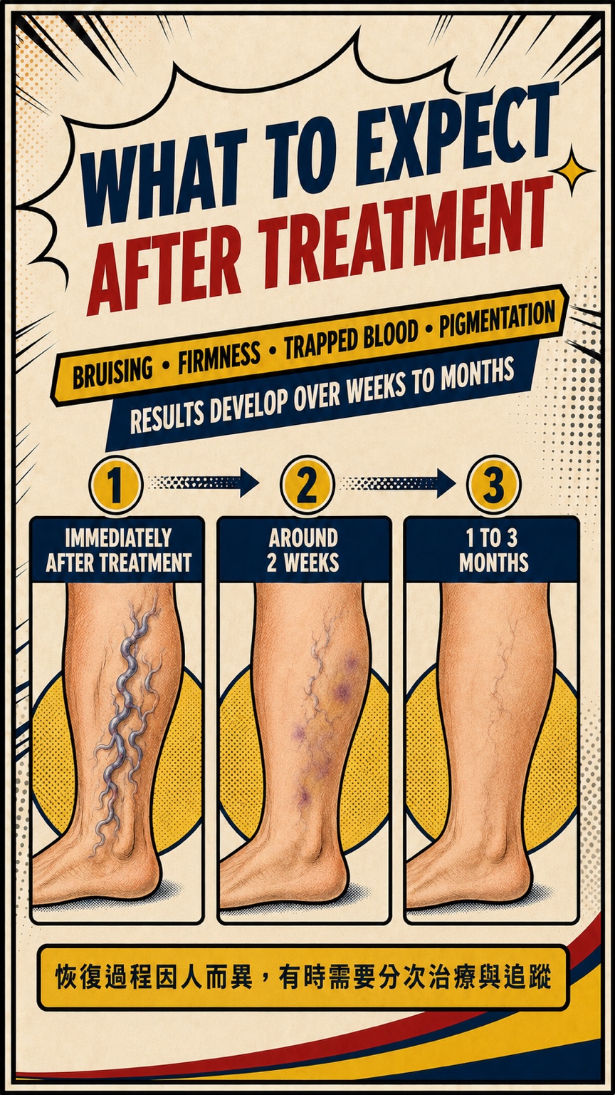
Why is the leg not instantly smooth after treatment?
- Temporary bruising, firmness, local tenderness, trapped blood or pigmentation may occur in treated areas.
- The vein and surrounding tissue need time to respond and be reabsorbed. Changes usually develop over weeks to months rather than being final on the treatment day.
- Recovery varies with vein size, treatment extent, method and individual factors. Staged treatment and follow-up may be needed.
Main-vein reflux and superficial bulging branches
| Comparison | Main-vein reflux | Superficial bulging branch |
|---|---|---|
| Location | Follows a more predictable superficial trunk | Close to the skin with a more variable course |
| Appearance | May not be directly visible | Often tortuous and rope-like |
| How it is confirmed | Standing ultrasound maps reflux | Clinical examination plus ultrasound tracing |
| Possible treatment | VenaSeal or another suitable method | Sclerotherapy, microphlebectomy or another individualized method |
Frequently asked questions
Does a bulging vein always mean that the great saphenous vein is refluxing?
No. A bulging vein is often a superficial tributary and may connect to the GSV, SSV, an accessory vein, a perforator or a local source. Standing ultrasound is needed to confirm the anatomy.
Is injecting only the visible veins enough?
It depends on the reflux source and vein size. If upstream main-vein reflux is present, treating surface branches alone may be incomplete. If the main trunks are competent, targeted branch treatment may be considered. Sclerotherapy still requires an individual assessment.
Are bruising, firmness or pigmentation normal after treatment?
These temporary changes can occur and often improve with time. Prompt review is appropriate if pain or swelling is worsening, the area becomes red and hot, or other unusual symptoms develop.
How long does it take to see results?
Appearance and symptoms generally evolve over weeks to months. Timing varies with vein size, treatment extent, method and individual factors, and staged treatment may be required.
Note: Seek urgent care for sudden one-sided leg swelling or pain, redness and heat, persistent bleeding from a varicose vein, chest pain or shortness of breath.
References
- De Maeseneer MG, Kakkos SK, Aherne T, et al. European Society for Vascular Surgery (ESVS) 2022 Clinical Practice Guidelines on the Management of Chronic Venous Disease of the Lower Limbs. Eur J Vasc Endovasc Surg. 2022;63(2):184–267. doi:10.1016/j.ejvs.2021.12.024. View source
- Gloviczki P, Lawrence PF, Wasan SM, et al. The 2022 Society for Vascular Surgery, American Venous Forum, and American Vein and Lymphatic Society clinical practice guidelines for the management of varicose veins of the lower extremities. Part I. J Vasc Surg Venous Lymphat Disord. 2023;11(2):231–261.e6. doi:10.1016/j.jvsv.2022.09.004. View source
- Gloviczki P, Lawrence PF, Wasan SM, et al. The 2023 Society for Vascular Surgery, American Venous Forum, and American Vein and Lymphatic Society clinical practice guidelines for the management of varicose veins of the lower extremities. Part II. J Vasc Surg Venous Lymphat Disord. 2024;12(1):101670. doi:10.1016/j.jvsv.2023.08.011. View source
- Morrison N, Gibson K, Vasquez M, Weiss R, Jones A. Five-year extension study of patients from a randomized clinical trial (VeClose) comparing cyanoacrylate closure versus radiofrequency ablation for the treatment of incompetent great saphenous veins. J Vasc Surg Venous Lymphat Disord. 2020;8(6):978–989. doi:10.1016/j.jvsv.2019.12.080. View source
This article and its illustrations provide general health education. They do not mean that every patient needs VenaSeal or sclerotherapy, and they do not guarantee a specific result. The source of reflux, treatment, recovery and outcome require individual clinical and standing-ultrasound assessment.
Dr LiangVascular
Begin with a careful assessment.
Symptoms, examination and ultrasound findings are considered before treatment options are discussed.
Book an Appointment