中文版
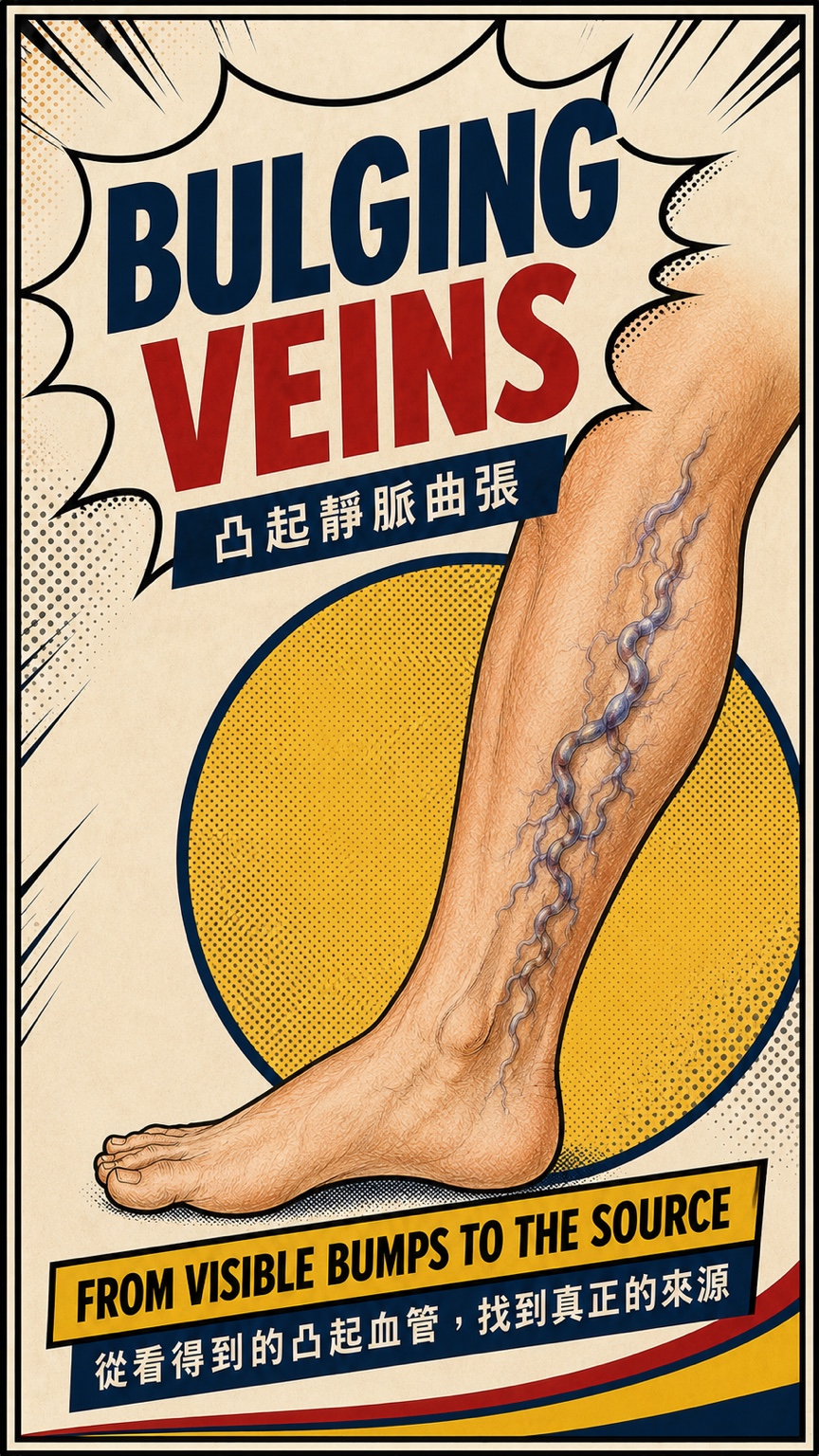
肉眼看到的凸起血管通常是靠近皮膚的表淺分支,卻不一定就是逆流的源頭。治療前先以站立式下肢靜脈超音波追蹤血流方向,才能分辨問題來自主幹、附屬靜脈、穿通枝,或較局部的分支。
- 外觀看起來相似的凸起血管,逆流來源可能完全不同
- 站立式超音波用來確認血流方向、來源與影響範圍
- 治療方式應依症狀、血管分布與超音波結果個別規劃
凸起血管不只影響外觀
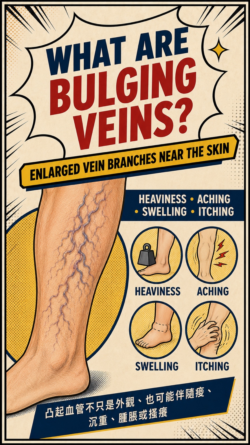
- 凸起靜脈通常是靠近皮膚、已擴張且變得彎曲的表淺分支。
- 有些人沒有明顯不舒服;有些人會出現腿部沉重、痠痛、腫脹、搔癢或久站後症狀加重。
- 若腳踝皮膚逐漸變色、變硬、反覆發炎或出現不易癒合的傷口,應儘早接受評估。
為什麼要先找逆流來源?
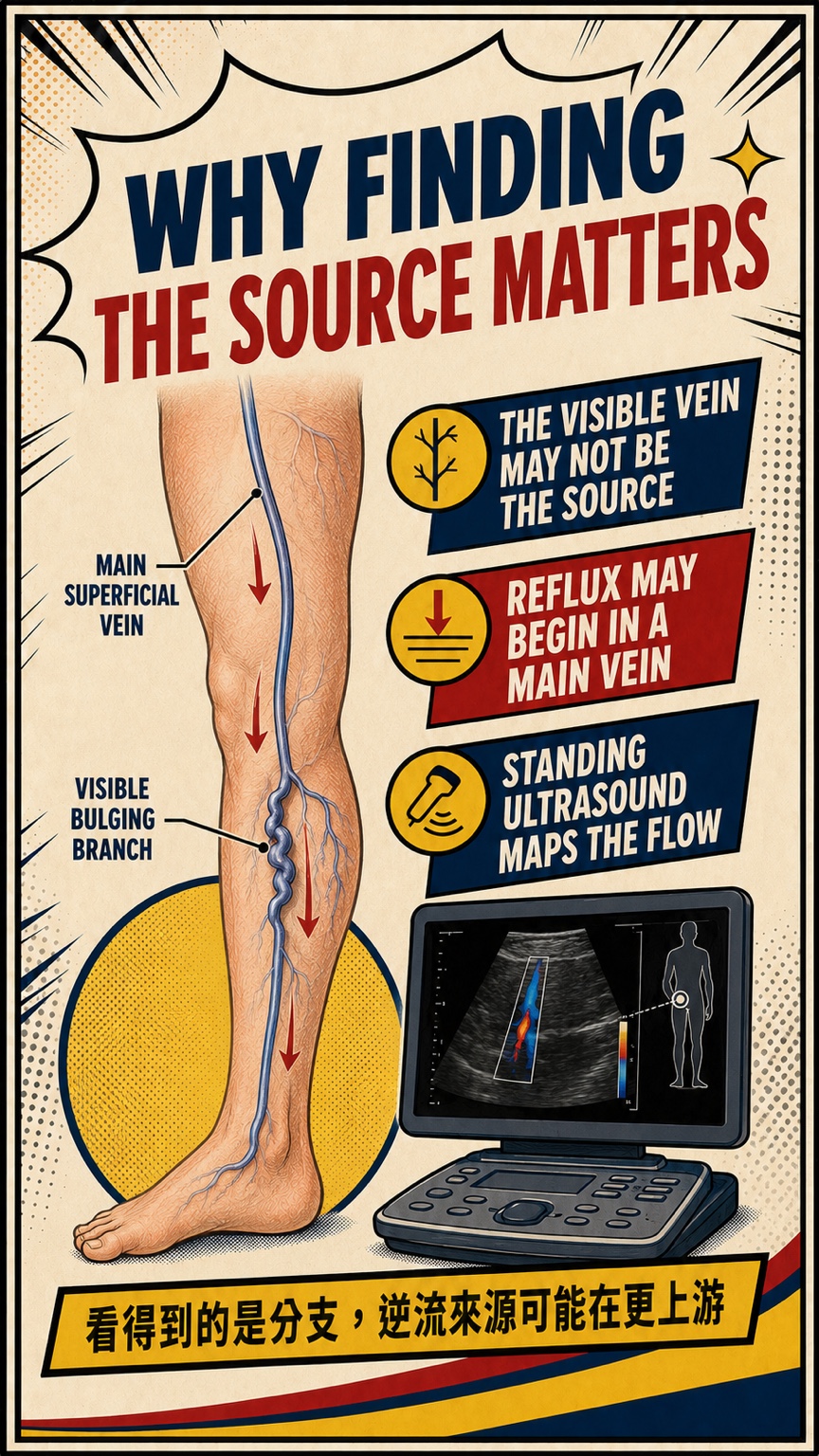
- 看得到的血管可能只是下游分支,逆流可能從較上游的主幹、附屬靜脈或穿通枝開始。
- 也有些人的主幹功能正常,問題主要位於局部表淺分支,因此不能只依外觀預設治療方式。
- 站立式超音波可以追蹤血流方向、逆流起點與範圍,建立每一條腿自己的血管地圖。
我會怎麼評估?
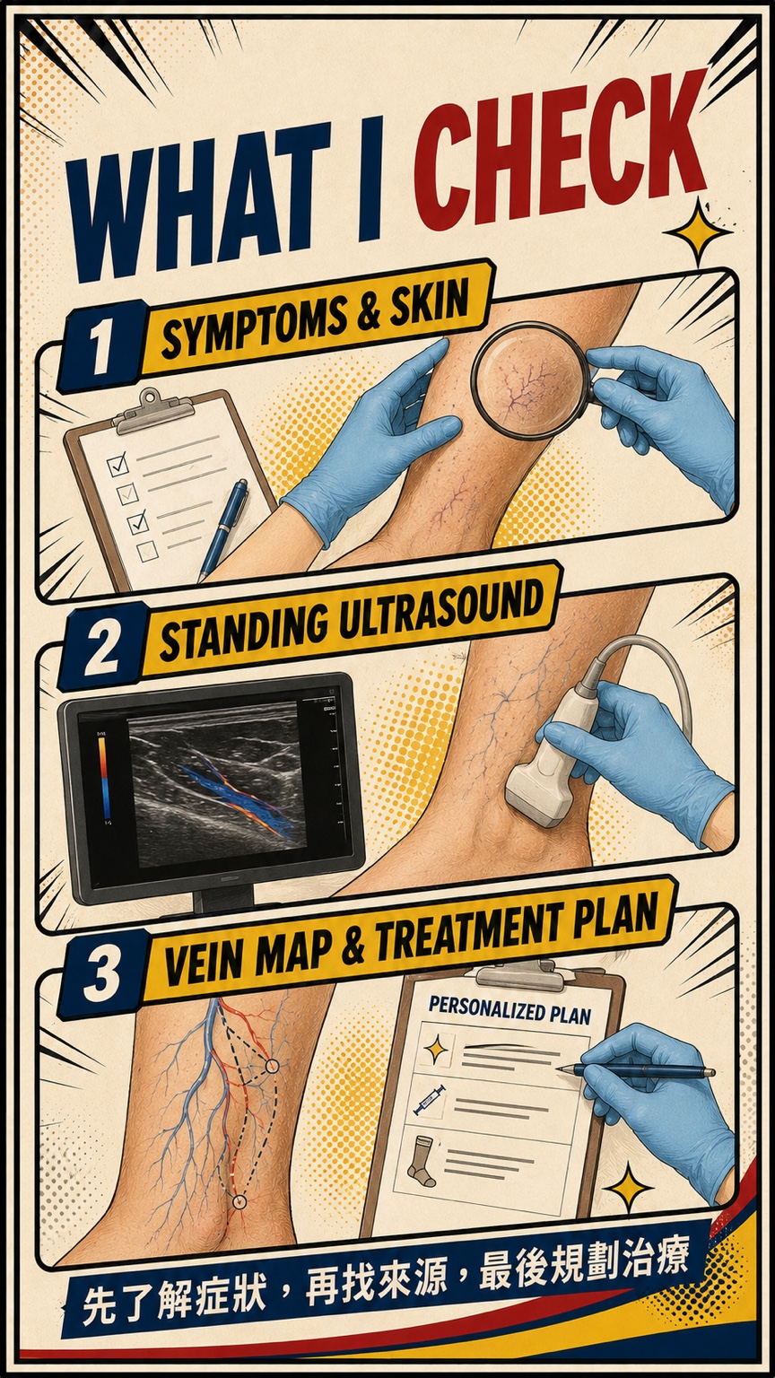
- 先了解最困擾的症狀、出現時間、久站影響、過去治療與皮膚變化。
- 需要時進行站立式下肢靜脈超音波,檢查表淺與深部靜脈,並追蹤逆流來源。
- 最後把症狀、外觀、血管走向與超音波結果放在一起,討論適合的治療順序與合理期待。
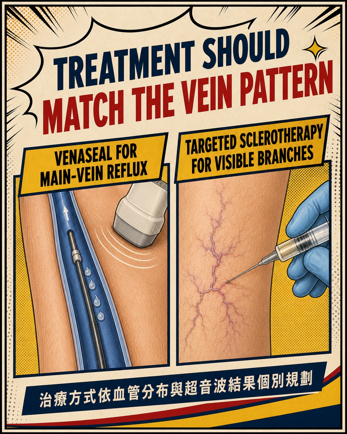
治療要配合血管分布
- 若有適合處理的主幹逆流,可依血管條件、過敏史與個人需求評估VenaSeal醫療膠或其他治療。
- 肉眼可見的凸起分支,可能安排超音波導引硬化劑注射、微創靜脈摘除或其他適合方式。
- 主幹與分支可以視情況同時處理,也可能分階段完成;並非每位病人都需要VenaSeal,也不是每條凸起血管都適合直接注射。
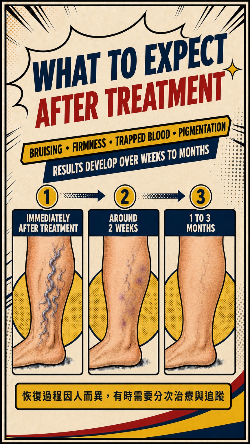
治療後為什麼不會立刻完全平整?
- 治療區可能暫時出現瘀青、硬塊、局部壓痛、血液滯留或色素沉著。
- 血管與周圍組織需要時間逐步反應和吸收,外觀變化通常以數週至數月觀察,不是治療當天立即定案。
- 恢復速度因血管大小、範圍、治療方式與個人體質而異,有時需要分次治療與追蹤。
主幹逆流與表淺凸起分支
| 比較 | 主幹逆流 | 表淺凸起分支 |
|---|---|---|
| 位置 | 沿較固定的表淺主幹走向 | 靠近皮膚,走向較多變 |
| 外觀 | 不一定直接看得到 | 常呈彎曲、繩索狀凸起 |
| 如何確認 | 站立式超音波追蹤逆流 | 外觀檢查並以超音波追查來源 |
| 可能的治療 | VenaSeal醫療膠或其他適合方式 | 硬化劑、微創摘除或其他個別方式 |
常見問題
腿上有凸起血管,就代表大隱靜脈一定逆流嗎?
不一定。凸起血管常是表淺分支,可能連接大隱靜脈、小隱靜脈、附屬靜脈、穿通枝或局部來源,需要以站立式超音波確認。
只把看得到的凸起血管打硬化劑就可以嗎?
要看逆流來源與血管大小。如果上游有主幹逆流,只處理表面分支未必完整;若主幹功能正常,則可針對局部分支規劃。硬化劑是否適合仍需個別評估。
治療後出現瘀青、硬塊或色素沉著正常嗎?
治療後可能暫時出現這些變化,通常會隨時間改善;但若疼痛或腫脹明顯加劇、發紅發熱,或有其他不尋常症狀,應儘快回診。
多久可以看到結果?
外觀與症狀通常需要數週至數月逐步觀察。實際時間取決於血管大小、治療範圍、治療方式與個人體質,有時需要分次處理。
提醒:如果單側腿突然腫痛、發紅發熱,曲張血管持續出血,或出現胸痛、呼吸困難,請立即就醫。
English
The veins you can see are often superficial tributaries, but they are not necessarily the source of reflux. Standing leg-vein ultrasound traces blood-flow direction and helps determine whether reflux arises from a main trunk, accessory vein, perforator or a more local branch.
- Similar-looking bulging veins can have very different sources of reflux
- Standing ultrasound maps blood-flow direction, source and extent
- Treatment should be planned according to symptoms, anatomy and ultrasound findings
Bulging veins are more than an appearance issue
- Bulging varicose veins are enlarged, tortuous superficial branches located close to the skin.
- Some people have no significant discomfort, while others experience heaviness, aching, swelling, itching or symptoms that worsen after standing.
- Progressive ankle discoloration, firmness, recurrent inflammation or a slow-healing wound deserves earlier assessment.
Why does finding the source matter?
- The visible vein may be a downstream branch while reflux begins in a main trunk, accessory vein or perforator further upstream.
- In other people, the main trunks remain competent and the problem is mainly local. Appearance alone cannot determine the right treatment.
- Standing ultrasound traces the direction, starting point and extent of reflux to create an individual vein map.
What do I check?
- We first discuss the symptoms that matter most, their timing, the effect of prolonged standing, previous treatment and skin changes.
- When appropriate, standing leg-vein ultrasound assesses the superficial and deep veins and traces the source of reflux.
- Symptoms, appearance, venous anatomy and ultrasound findings are then combined into a treatment sequence with realistic expectations.
Treatment should match the vein pattern
- When suitable main-vein reflux is present, VenaSeal medical adhesive or another treatment may be considered according to anatomy, allergy history and personal needs.
- Visible bulging tributaries may be treated with ultrasound-guided sclerotherapy, microphlebectomy or another appropriate method.
- Trunks and branches may be treated together or in stages. Not every patient needs VenaSeal, and not every bulging vein is suitable for direct injection.
Why is the leg not instantly smooth after treatment?
- Temporary bruising, firmness, local tenderness, trapped blood or pigmentation may occur in treated areas.
- The vein and surrounding tissue need time to respond and be reabsorbed. Changes usually develop over weeks to months rather than being final on the treatment day.
- Recovery varies with vein size, treatment extent, method and individual factors. Staged treatment and follow-up may be needed.
Main-vein reflux and superficial bulging branches
| Comparison | Main-vein reflux | Superficial bulging branch |
|---|---|---|
| Location | Follows a more predictable superficial trunk | Close to the skin with a more variable course |
| Appearance | May not be directly visible | Often tortuous and rope-like |
| How it is confirmed | Standing ultrasound maps reflux | Clinical examination plus ultrasound tracing |
| Possible treatment | VenaSeal or another suitable method | Sclerotherapy, microphlebectomy or another individualized method |
Frequently asked questions
Does a bulging vein always mean that the great saphenous vein is refluxing?
No. A bulging vein is often a superficial tributary and may connect to the GSV, SSV, an accessory vein, a perforator or a local source. Standing ultrasound is needed to confirm the anatomy.
Is injecting only the visible veins enough?
It depends on the reflux source and vein size. If upstream main-vein reflux is present, treating surface branches alone may be incomplete. If the main trunks are competent, targeted branch treatment may be considered. Sclerotherapy still requires an individual assessment.
Are bruising, firmness or pigmentation normal after treatment?
These temporary changes can occur and often improve with time. Prompt review is appropriate if pain or swelling is worsening, the area becomes red and hot, or other unusual symptoms develop.
How long does it take to see results?
Appearance and symptoms generally evolve over weeks to months. Timing varies with vein size, treatment extent, method and individual factors, and staged treatment may be required.
Note: Seek urgent care for sudden one-sided leg swelling or pain, redness and heat, persistent bleeding from a varicose vein, chest pain or shortness of breath.
References
- De Maeseneer MG, Kakkos SK, Aherne T, et al. European Society for Vascular Surgery (ESVS) 2022 Clinical Practice Guidelines on the Management of Chronic Venous Disease of the Lower Limbs. Eur J Vasc Endovasc Surg. 2022;63(2):184–267. doi:10.1016/j.ejvs.2021.12.024. 查看來源
- Gloviczki P, Lawrence PF, Wasan SM, et al. The 2022 Society for Vascular Surgery, American Venous Forum, and American Vein and Lymphatic Society clinical practice guidelines for the management of varicose veins of the lower extremities. Part I. J Vasc Surg Venous Lymphat Disord. 2023;11(2):231–261.e6. doi:10.1016/j.jvsv.2022.09.004. 查看來源
- Gloviczki P, Lawrence PF, Wasan SM, et al. The 2023 Society for Vascular Surgery, American Venous Forum, and American Vein and Lymphatic Society clinical practice guidelines for the management of varicose veins of the lower extremities. Part II. J Vasc Surg Venous Lymphat Disord. 2024;12(1):101670. doi:10.1016/j.jvsv.2023.08.011. 查看來源
- Morrison N, Gibson K, Vasquez M, Weiss R, Jones A. Five-year extension study of patients from a randomized clinical trial (VeClose) comparing cyanoacrylate closure versus radiofrequency ablation for the treatment of incompetent great saphenous veins. J Vasc Surg Venous Lymphat Disord. 2020;8(6):978–989. doi:10.1016/j.jvsv.2019.12.080. 查看來源
本文與圖像提供一般衛教,不代表每位病人都需要VenaSeal或硬化劑治療,也不保證特定療效。實際逆流來源、治療方式、恢復時間與結果需依症狀、站立式超音波及個人狀況評估。
Dr LiangVascular
從完整評估開始
先整合症狀、理學檢查與超音波結果,再討論適合的治療選擇。
預約門診