中文版
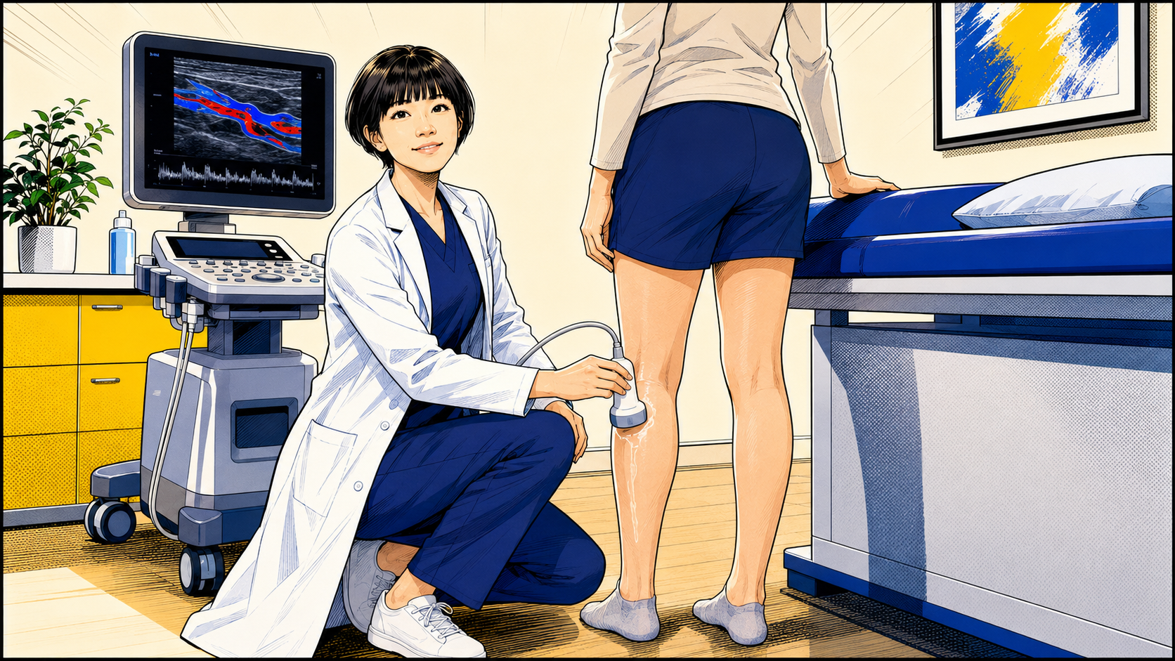
檢查不需要打針,也沒有輻射。醫師會依臨床情況在站立或其他讓靜脈充分充盈的姿勢下移動探頭,必要時輕壓小腿或使用其他方式觀察血液流動。
- 在臨床適當時找出逆流位置、方向與範圍
- 評估表淺與深部靜脈,並建立血管地圖
- 協助診斷與治療規劃,但不能單獨決定是否治療
哪些情況可能需要站立式靜脈超音波?
- 有痠痛、沉重、腫脹、搔癢等症狀的靜脈曲張,或曾治療後再次出現的曲張血管。
- 腿部腫脹、皮膚變色或變硬、傷口癒合不佳,且臨床上懷疑可能與靜脈疾病有關。
- 正在規劃治療,需要了解靜脈解剖、逆流來源與不同血管之間的連接。
- 治療後仍有症狀、外觀疑慮,或出現新的血管變化,需要分辨殘存、復發或新的逆流模式。
哪些情況未必需要例行完整檢查?
- 只有少量、無症狀且純外觀性的表淺細小血管,不代表一定需要例行做完整 duplex ultrasound。
- 若合併腿部症狀、腫脹、皮膚變化、較粗的曲張血管、復發性血管或其他臨床疑慮,檢查價值會不同。
- 是否安排檢查仍由病史、症狀、臨床檢查與患者想處理的問題共同決定。
檢查會看到什麼?
- 表淺與深部靜脈的解剖、通暢情況與血流方向。
- 逆流的位置、方向、範圍,以及相關血管的連接模式。
- 過去治療後是否有殘存、復發或新出現的逆流。
- 需要治療規劃時,可用血管地圖協助判斷應處理的來源與範圍。
超音波不能單獨決定什麼?
- 單一血管直徑或孤立的逆流發現,不能單獨決定是否需要治療。
- 超音波看到逆流,不代表所有腿痛、腿腫或其他不適都一定由該血管造成。
- 症狀、理學檢查、靜脈解剖、逆流模式、皮膚變化、生活影響與個別需求,必須一起解讀。
檢查前要準備嗎?
- 通常不需要空腹,也沒有輻射或顯影劑。
- 穿方便露出大腿與小腿的衣物,並帶上過去的超音波、手術或治療資料。
- 可以先記下症狀出現的位置、時間、與久站或活動的關係,以及最想了解的問題。
只看外觀與超音波評估的差別
| 比較 | 只看腿上血管 | 站立式超音波 |
|---|---|---|
| 能看到什麼 | 表面血管外觀 | 表淺與深部靜脈、血流方向與逆流模式 |
| 能否找逆流來源 | 無法可靠判斷 | 可在臨床適當時定位並建立血管地圖 |
| 治療規劃 | 可能只注意看得到的血管 | 協助整合解剖、來源與處理範圍 |
| 檢查方式 | 肉眼與理學檢查 | 非侵入性、無輻射,通常不需打針 |
常見問題
第一次看血管外科,一定會做站立式超音波嗎?
不一定。醫師會先了解病史、症狀與臨床檢查;當懷疑症狀可能與靜脈有關,或需要確認逆流來源、復發模式或治療規劃時,才依需要安排檢查。
為什麼靜脈超音波常在站立或下垂姿勢評估?
讓腿部靜脈在重力影響下充分充盈,有助於觀察血流與逆流模式。實際姿勢仍會依安全、患者狀況與檢查目的調整。
血管較粗或超音波看到逆流,就一定要治療嗎?
不一定。單一血管直徑或孤立的逆流發現不能單獨決定治療;症狀、檢查、解剖、逆流模式、皮膚變化、生活影響與個別需求必須一起評估。
提醒:不是每一條小青筋都需要完整超音波,也不能只靠單一超音波數字決定治療;是否檢查與如何處理,要回到症狀、臨床發現與個別需求。
English
The examination is non-invasive and uses no radiation or injections. According to the clinical situation, the veins are assessed while standing or in another dependent position that allows adequate venous filling, with manoeuvres used to observe blood flow.
- Maps the location, direction and extent of reflux when clinically appropriate
- Assesses the superficial and deep venous systems
- Supports diagnosis and treatment planning but does not decide treatment by itself
When might standing venous duplex ultrasound be needed?
- Symptomatic varicose veins, including aching, heaviness, swelling or itching, and varicose veins that have appeared again after previous treatment.
- Swelling, skin discoloration or induration, or a wound that is not healing normally when venous disease is clinically suspected.
- Treatment planning when venous anatomy, the source of reflux and connections between vessels need clarification.
- Persistent symptoms, appearance concerns or new venous changes after previous treatment, when residual, recurrent or newly developed reflux needs to be distinguished.
When might a routine full scan not be necessary?
- A small number of asymptomatic superficial vessels that are purely cosmetic does not automatically require a routine full duplex examination.
- The value of scanning changes when there are associated symptoms, swelling, skin changes, larger varicose veins, recurrent vessels or broader clinical concerns.
- The decision remains based on the history, symptoms, examination and the problem the patient wants assessed.
What does the examination show?
- The anatomy, patency and blood-flow direction of the superficial and deep venous systems.
- The location, direction and extent of reflux, and the pattern of connections between related veins.
- Residual, recurrent or newly developed reflux after previous treatment.
- A venous map that can support treatment planning when intervention is being considered.
What can ultrasound not decide by itself?
- A vein diameter or an isolated reflux finding should not by itself determine whether treatment is necessary.
- Reflux on ultrasound does not prove that every episode of leg pain, swelling or other discomfort is caused by that vein.
- Symptoms, examination, anatomy, reflux pattern, skin changes, impact on daily life and individual circumstances must be interpreted together.
How should I prepare?
- Fasting is usually unnecessary, and the examination uses no radiation or contrast injection.
- Wear clothing that allows the thigh and lower leg to be exposed, and bring previous ultrasound, procedure or treatment records.
- Note where and when symptoms occur, whether they relate to standing or activity, and the questions you most want answered.
Appearance alone compared with duplex assessment
| Comparison | Looking at the leg | Standing duplex ultrasound |
|---|---|---|
| Information | Surface appearance | Superficial and deep veins, blood-flow direction and reflux pattern |
| Source of reflux | Cannot be identified reliably | Can be mapped when clinically appropriate |
| Treatment planning | May focus only on visible vessels | Helps integrate anatomy, source and treatment extent |
| Examination | Visual and physical examination | Non-invasive, no radiation and usually no injection |
Frequently asked questions
Does every first vascular appointment include standing duplex ultrasound?
No. The history, symptoms and clinical examination come first. Duplex is arranged when a venous cause is suspected or when reflux mapping, recurrent-disease assessment or treatment planning is clinically useful.
Why is venous ultrasound often performed while standing or in a dependent position?
Allowing the leg veins to fill under gravity helps the examiner observe blood flow and reflux. The exact position is adjusted for safety, the patient's condition and the purpose of the examination.
Does a larger vein or reflux on ultrasound mean treatment is necessary?
No. A vein diameter or isolated reflux finding should not determine treatment by itself. Symptoms, examination, anatomy, reflux pattern, skin changes, impact on daily life and individual circumstances must be considered together.
Note: Not every small visible vein requires a full duplex scan, and treatment should not be decided from a single ultrasound measurement. Symptoms, clinical findings and individual circumstances remain essential.
References
- Gloviczki P, Lawrence PF, Wasan SM, et al. The 2022 Society for Vascular Surgery, American Venous Forum, and American Vein and Lymphatic Society Clinical Practice Guidelines for the Management of Varicose Veins of the Lower Extremities. Part I: Duplex Scanning and Treatment of Superficial Truncal Reflux. Journal of Vascular Surgery: Venous and Lymphatic Disorders. 2023;11(2):231–261.e6. 查看來源
- De Maeseneer MG, Kakkos SK, Aherne T, et al. European Society for Vascular Surgery 2022 Clinical Practice Guidelines on the Management of Chronic Venous Disease of the Lower Limbs. European Journal of Vascular and Endovascular Surgery. 2022;63(2):184–267. 查看來源
- National Institute for Health and Care Excellence. Varicose veins: diagnosis and management (CG168). Duplex ultrasound assessment recommendations. 查看來源
本文提供一般衛教,不取代個別診斷或治療。若有急性不舒服,請先就近就醫。
Dr LiangVascular
從完整評估開始
先整合症狀、理學檢查與超音波結果,再討論適合的治療選擇。
預約門診