中文版
不過,超音波看到血管狹窄,不代表一定要立刻做血管擴張。我通常會一起評估廔管的震顫、洗腎狀況與超音波結果,再決定是否需要處理。
- 原本明顯的震顫變弱,或摸起來和以前不同
- 洗腎下針越來越困難,經常需要重新穿刺
- 洗腎機器反覆出現壓力或血流警示
- 拔針後的止血時間比以前明顯變久
- 廔管手臂持續腫脹,或表面血管越來越突出
- 洗腎效率下降,無法達到原本設定的血流速度
哪些變化可能代表廔管不順?
- 震顫變弱、變得斷斷續續,或摸起來和平常不同。
- 洗腎下針越來越困難,或機器經常出現壓力、血流警示。
- 拔針後止血時間比平常明顯延長。
- 廔管手臂持續腫脹,或洗腎效率下降。
醫師會怎麼檢查?
- 先用手觸摸廔管,評估震顫、脈搏與血流聲。
- 用超音波查看狹窄的位置、血流速度,以及是否形成血栓。
- 一起確認上游與下游血管是否通暢,以及狹窄是否真的影響洗腎。
- 治療重點不是只看一個數字,而是找出真正造成問題的狹窄。
什麼情況可能需要血管擴張?
- 如果超音波確認明顯狹窄,同時出現洗腎血流不足、下針困難、止血變久或震顫改變,通常會考慮治療。
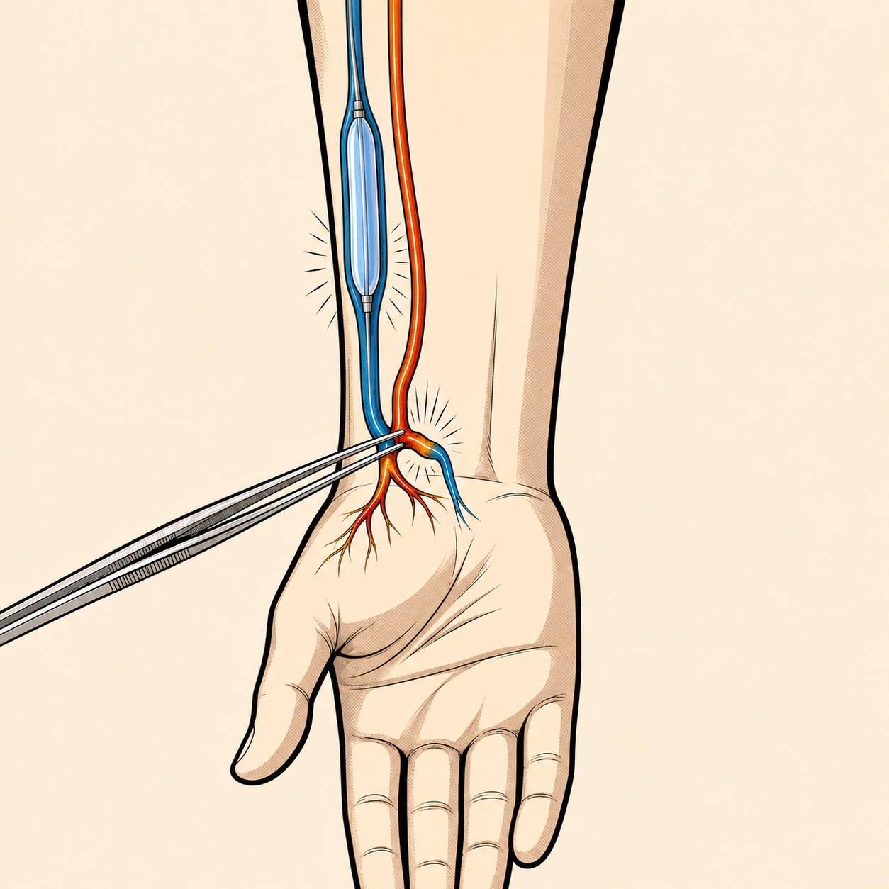
- 治療時會從血管放入細小導管,再用氣球把狹窄處撐開,改善廔管血流。
- 多數情況不需要開刀,通常使用局部麻醉。
血管擴張後就不會再變窄嗎?
- 血管擴張可以改善血流,但不能保證永遠不再狹窄。
- 部分廔管可能在一段時間後再次出現問題,因此仍要持續觀察震顫與洗腎狀況。
- 若同一位置反覆狹窄,醫師會再評估其他治療方式或手術重建。
廔管變窄時,一般會怎麼處理?
| 情況 | 一般處理方向 |
|---|---|
| 只有影像顯示狹窄,洗腎與震顫都穩定 | 持續觀察與追蹤 |
| 震顫改變、下針困難或止血變久 | 安排超音波與血管評估 |
| 狹窄已影響洗腎血流 | 考慮血管擴張 |
| 震顫完全消失 | 當天儘快處理,可能已形成血栓 |
提醒:每天花幾秒鐘摸摸廔管的震顫,熟悉它平常的感覺。如果震顫突然消失、手臂快速腫起來,或傷口持續大量出血,請不要等到下一次洗腎,應儘快聯絡洗腎室或安排血管外科評估。
English
However, a narrowing seen on ultrasound does not automatically mean that angioplasty is required. I consider the feel of the access, dialysis performance and ultrasound findings together before recommending treatment.
- The usual thrill feels weaker or different
- Needle insertion becomes difficult or requires repeated attempts
- The dialysis machine repeatedly shows pressure or blood-flow alarms
- Bleeding after needle removal takes noticeably longer to stop
- The access arm remains swollen or surface veins become more prominent
- Dialysis efficiency falls or the prescribed blood-flow rate cannot be achieved
What changes may suggest an access problem?
- The thrill becomes weaker, intermittent or different from usual.
- Needling becomes more difficult, or the machine repeatedly shows pressure or flow alarms.
- Bleeding after needle removal takes noticeably longer than usual.
- The access arm remains swollen or dialysis efficiency falls.
How is the access assessed?
- The access is examined by feeling its pulse and thrill and listening to the blood-flow sound.
- Ultrasound can assess the location of narrowing, blood flow and whether a clot is present.
- The inflow and outflow vessels are checked to determine whether the narrowing is affecting dialysis.
- The goal is not simply to identify a number on a scan, but to find the lesion causing the problem.
When may angioplasty be considered?
- Treatment may be considered when significant narrowing is confirmed together with reduced dialysis flow, difficult cannulation, prolonged bleeding or a change in the thrill.
- A small catheter is placed inside the vessel, and a balloon is inflated to open the narrowed area and improve flow.
- The procedure is generally performed under local anaesthesia and usually does not require open surgery.
Can narrowing return after treatment?
- Angioplasty can improve blood flow, but it cannot guarantee that the vessel will never narrow again.
- Continued monitoring of the thrill and dialysis performance remains important.
- Repeated narrowing may require another treatment strategy or surgical reconstruction.
How is a narrowed access usually managed?
| Situation | Usual next step |
|---|---|
| Narrowing on imaging, but dialysis and the thrill remain stable | Continue clinical monitoring |
| Change in thrill, difficult needling or prolonged bleeding | Arrange ultrasound and vascular assessment |
| Confirmed stenosis affecting dialysis flow | Consider angioplasty |
| Complete disappearance of the thrill | Seek same-day assessment for possible thrombosis |
Note: Take a few seconds each day to feel the usual vibration of your dialysis access. If the thrill suddenly disappears, the arm rapidly becomes swollen, or bleeding cannot be controlled, do not wait until the next dialysis session. Contact the dialysis unit or arrange urgent vascular assessment.
References
- Lok CE, et al. KDOQI Clinical Practice Guideline for Vascular Access: 2019 Update. Am J Kidney Dis. 2020;75(4 Suppl 2):S1–S164. 查看來源
- Schmidli J, et al. Vascular Access: 2018 Clinical Practice Guidelines of the European Society for Vascular Surgery. Eur J Vasc Endovasc Surg. 2018;55:757–818. 查看來源
- Sidawy AN, et al. The Society for Vascular Surgery clinical practice guidelines for arteriovenous hemodialysis access. J Vasc Surg. 2008;48(5 Suppl):2S–25S. 查看來源
本文提供一般衛教,不取代個別診斷或治療。若有急性不舒服,請先就近就醫。
Dr LiangVascular
從完整評估開始
先整合症狀、理學檢查與超音波結果,再討論適合的治療選擇。
預約門診